Sharui Shan,
Xuming Huang ![]() ,
Mingxing Zhang,
Yihua Shi
,
Mingxing Zhang,
Yihua Shi
For correspondence:- Xuming Huang Email: 781607917@qq.com Tel:+862061335054
Received: 10 January 2016 Accepted: 28 May 2016 Published: 28 June 2016
Citation: Shan S, Huang X, Zhang M, Shi Y. Anti-cancer and antioxidant properties of phenolics isolated from Toona sinensis A Juss acetone leaf extract. Trop J Pharm Res 2016; 15(6):1205-1213 doi: 10.4314/tjpr.v15i6.13
© 2016 The authors.
This is an Open Access article that uses a funding model which does not charge readers or their institutions for access and distributed under the terms of the Creative Commons Attribution License (http://creativecommons.org/licenses/by/4.0) and the Budapest Open Access Initiative (http://www.budapestopenaccessinitiative.org/read), which permit unrestricted use, distribution, and reproduction in any medium, provided the original work is properly credited..
Purpose: To investigate the antioxidant and anticancer activities of phenolics from the leaf extract of Toona sinensis (TS).
Methods: Acetone leaf extract of TS was screened for total phenolic and flavanoid contents, and the flanonoids were subjected to high performance liquid chromatographic (HPLC) analysis. Antioxidant properties were assessed via oxygen radical absorbance capacity (ORAC), peroxyl radical scavenging capacity (PSC) and cellular antioxidant activity (CAA), while anti-proliferative activity ins HepG2 cell line was assessed using methylene blue assay.
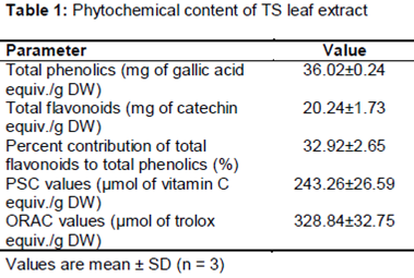
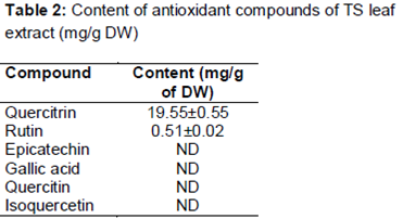
Results: The extract contained 36.02 ± 0.24 mg of gallic acid equiv/g dry weight (DW) and 20.24 ± 1.73 mg of catechin equiv/g DW of total phenolic and total flavonoid, respectively. The levels of rutin and quercitrin were 0.51 and 19.55 mg/g, respectively. Epicatechin, gallic acid, quercitin, isoquercetin were not detected. The extract showed significant antioxidant potential and high anti-proliferation capacity with low cytotoxicity against HepG2 cell in vitro. The underlying mechanism of anti-proliferative effect was induction of apoptosis.
Conclusion: TS leaf extract possesses significant in vitro antioxidant properties and anti-proliferative effect against HepG2 cells, which make it a potential anticancer drug source.
Introduction
Phytochemicals are plant-based non-nutrient secondary metabolites, which provide multiple positive health effect. These metabolites are increasingly recognized as efficacious components in the prevention and management of chronic disease, such as cancer, diabetes, and cardiovascular diseases [1].
Toona sinensis A. Juss is a deciduous tree which belongs to the family Meliaceae. All parts of TS are medicinally useful. Indeed the plant possesses many biological functions such as antitumor activity [2], antioxidant capacity [3], and anti-inflammatory properties [4]. It is also used for treating severe acute respiratory syndrome [5], and for improving hyperglycemia and dyslipidemia [6]. The young leaves of TS are popular in traditional Chinese medicines and have been used for long as a safe vegetable source [7]. Non-polar extracts of TS leaves contain constituents that can prevent type-2 diabetes and hepatosteatosis [8]. Previous studies showed that TS leaves possess anti-oxidative activity and anti-inflammatory activities [9]. TS is used for treatment of liver cancer in Gansu province of China [10]. Although the anti-diabetic, antioxidant and anti-inflammatory activities of TS leaves have been reported, there is lack of information on their phenolic profiles, cellular antioxidant activity and in vitro anti-proliferative activity.
Methods
Chemicals and materials
2,2ˊ-Azobis-amidino-propane (ABAP), catechin hydrate, gallic acid, Folin-Ciocalteu reagent and dichlorofluorescin diacetate (DCFH-DA) were obtained from Sigma Chemical Co. (St. Louis, MO, US). All solvents used were of analytical grade. Williams medium E (WME), insulin, and other cell culture media were purchased from Gibco U.S. Biotechnology Co. Fetal bovine serum (FBS) was bought from Tianhang Biotech Co. Inc. (Zhejiang, China). HepG2 cell line was obtained from the Sun Yat-sen University Culture Collection.
Samples treatment and preparation
One kilogram of the leaves was collected from Anhui-China in spring 2014. The leaves were washed with distilled water to remove dirt, followed by freezing at -80 °C for 24 h and vacuum freeze-drying. The dried leaves were ground into powders (60 mesh) and stored at -80 °C prior to analysis.
Extraction procedures
The powder extraction procedure was a modification of the classical method described by Chu et al [11]. The powder (10 g) was homogenized with chilled acetone (80 %) for 5 min. The homogenate was filtered under vacuum, and the residue was re-extracted and filtered three times. The extracts were pooled in flasks and evaporated at 45 °C until no acetone was left. The residue was dissolved in a little volume of distilled water and stored at -40 °C until used.
Estimation of total phenolic content
Total phenolic content was determined as gallic acid equivalent using gallic acid standard curve [12], and the final value was expressed as milligram of gallic acid equivalent (GAE) per gram DW.
Measurement of total flavonoid content
The total flavonoid content was determined using the sodium borohydride/chloranil-based method as described earlier [13]. Values were calculated using catechin hydrate standard curve and expressed as milligram of catechin equivalent (CE)/g DW. Data are showed as mean ± SD.
Analysis of flavonoids by HPLC
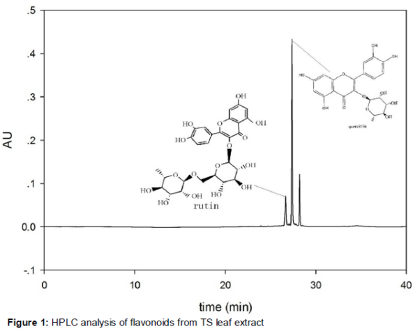
Flavonoid compounds in the sample extract were quantified using HPLC. HPLC system was assembled with Waters Associates chromatography (Waters Corp, Milford, US), and spectrophotometer set at 360 nm was used. A Sun Fire C18 phase column (5 μm particle size, 250 mm × 4.6 mm) was employed for chromatographic separation at 30 °C. The binary mobile phase consisted of methanol (A) and water (B). After injection of 10 μL sample, the system was eluted with 5 % A for 5 min; then increased to 10, 15, 20 and 45 % each at 5-min intervals. Then the gradient solvent A was increased to 100 % in 5 min, kept at 100 % for 10 min, and recycled back to 5 % after 5 min, and kept for 5 min prior to running the next sample. All process flow rate was 1.0 mL/min. The peaks were identified through their retention times by comparing with UV-visible spectra of standards.
Antioxidant assays
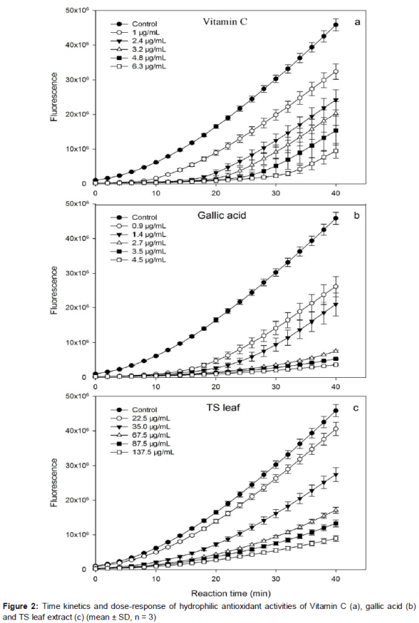
Total antioxidant activities were determined by hydrophilic peroxyl radical scavenging capacity assay and oxygen radical scavenging capacity assay. Hydrophilic peroxyl radical scavenging capacity of the extract was evaluated using the method as outlined previously [14]. In a run, pure compounds or sample extract appropriately diluted in phosphate buffer were transferred into reaction cells on a 96-well plate followed by the addition of DCFH. The reaction was initiated by adding ABAP and read at once. Each set of dilutions and control was added in triplicate in 96-well plate. Fluorescence was monitored at 485 nm excitation and 535 nm emission in a fluorescent spectrophotometer (SoftMax systems, Molecular Devices, US). Result was presented as micromoles of vitamin C equivalent (VCE) per gram of sample.
Oxygen radical scavenging capacity was measured as described by Prior et al [15,16], using black, clear-bottom, 96-well microplates. Extract diluent and trolox standard were added to appropriate wells and incubated at 37 °C for 10 min. Fluoroscein working solution was added to each well and incubated at 37 °C for 20 min. After incubation, freshly prepared ABAP was added to each well (except the control well) and the wells were immediately read in Fluoroskan Ascent FL plate-reader at excitation of 485 nm and emission of 535 nm for 35 cycles every 4.5 min. ORAC value was expressed as mean micromoles of trolox equivalent (TE) per g DW.
Cell culture
HepG2 cells were cultured in WME, containing 2 mM L-glutamine, 10 mM Hepes, 5 μg/mL insulin, 0.05 μg/mL hydrocortisone, 50 μg/mL streptomycin, 50 units/mL penicillin, 5 % FBS and 100 μg/mL gentamicin. Cells were incubated at 37 °C with 5 % CO2, and seeded/sub-cultured when in exponential growth phase.
Cytotoxicity of extract against HepG2
Cytotoxicity was measured as described formerly [17]. The concentration of extract that decreased cell numbers by 10 % when compared to the control was considered to be cytotoxic.
Cellular antioxidant activity (CAA)
The cellular antixoidant activity of the extract was determined following the method of Wolfe et al [18]. HepG2 cells were seeded into black, clear-bottom, 96-well microplate. Cell medium was removed after 24 h of seeding, and the cells were washed with PBS, and treated in triplicate with solutions containing different concentrations of quercetin or extracts plus DCFH-DA dissolved in antioxidant treatment medium for 1 h. If a PBS wash was to be utilized, wells were washed with PBS followed by addition of ABAP solution. The microplate was read in a Fluoroskan Ascent FL plate-reader at excitation of 485 nm and emission of 535 nm for 13 cycles every 5 min. The area under the fluorescence-versus-time curve was calculated to determine CAA value. EC50 values were converted to CAA values, which were expressed as micromoles of quercetin equivalent (QE) per 100 g of sample, using the mean EC50 value for quercetin from three separate experiments.
Inhibition of cell proliferation
The anti-proliferation effects of the extract were determined using cell methylene blue colorimetric assay [17]. The anti-proliferative effects were assessed by the EC50 values, expressed as milligrams of sample extract per milliliter.
Assessment of apoptosis by flow cytometry
The apoptosis of HepG2 cell was analyzed by flow cytometer employing an annexin V-FITC and PI apoptosis detection kit. In brief, the cells were treated with 0, 4, 6 and 8 mg/mL of extract for 24 h followed by harvesting, washing and incubation with annexin V-FITC and PI at room temperature for 20 min. Aluminum foil was used during all staining processes to avoid light and; the cells were analyzed by flow cytometer.
Statistical analysis
Statistical analysis was performed using SPSS software 13.0 (SPSS Inc., Chicago, IL, USA), while dose-effect analysis was done using Calcusyn software version 2.0 (Biosoft, Cambridge, UK). All data are reported as mean ± SD (n = 3).
Results
Total phenolic and flavonoid contents
Measured values of total phenolic and flavonoid contents are given in . The total phenolic content of the leaf extract of TS was 36.02 mg of GCE/g DW, which was less than reported level of polyphenolic content (79.90 mg of GCE/g) in TS leaf [19]. However, this value was 45.28 times and 98.25 times higher compared to spinach and cabbage, respectively [11]. This indicated that TS leaf is a good source of phenolics.
Total flavonoid content of TS leaves extract was 20.24 ± 1.73 mg of CE/g DW. This amount was relatively higher than the value reported earlier (12.92 ± 0.95 mg of CE/g sample) in the aqueous extract of TS leaf [20]. In the studied sample, contribution of total flavonoids to total phenolics was 32.92 ± 2.65 %.
Flavonoid compounds
Flavonoids such as quercitrin, rutin, epicatechin, gallic acid, quercitin and isoquercetin were identified and quantified by comparing retention times with those of authentic standards using HPLC method. Only two compounds quercitrin and rutin were detected and quantified at concentrations of 19.55 mg/g DW (1.95 %) and 0.51 mg/g DW (0.51 %), respectively (, ). Epicatechin, gallic acid, quercitin, and isoquercetin were below the detection limit.
Total in vitro antioxidant activity
Reactive oxygen species (ROS) have been reported as the causative factor in many chronic diseases. Oxidative stress caused by ROS can be prevented by natural antioxidant compounds [21]. The results of in vitro antioxidant activities of the extract are displayed in and . Although antioxidant activities of plant extracts can be determined by various methods, PSC and ORAC methods are the latest and more authentic techniques. PSC assay incorporates DCFH-DA as a fluorescent probe, and is suitable for analyzing both hydrophilic and lipophilic antioxidants. This method is reliable, sensitive, rapid, precise, and reproducible [22]. The PSC value was 243.26 ± 26.59 μmol VCE/g DW ( and ). Food and pharmaceutical industries usually use oxygen radical absorbance capacity (ORAC) assay to test total antioxidant activity. In a typical ORAC assay, the loss of fluorescence of B-phycoerythrin is an indication of the degree of damage from its reaction with the peroxyl radical [16]. The antioxidant activity is valued by assessing the area under the fluorescence decay curve of the extract relative to that of the blank containing PBS [14]. The TS leaf extract had an ORAC value 328.84 ± 32.75 μmol TE/g DW ().
Cellular antioxidant activity
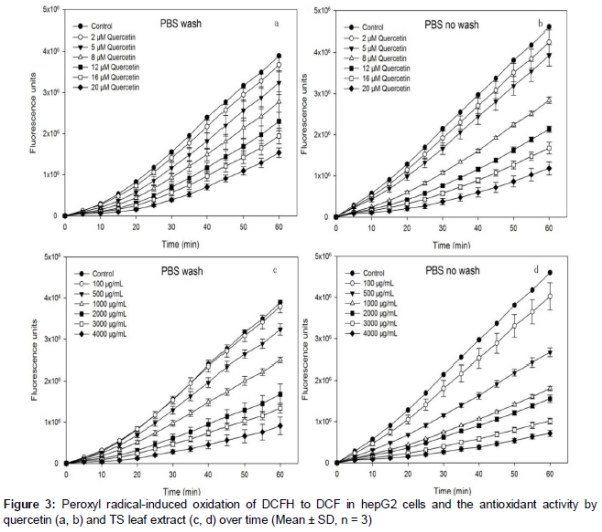
The kinetic of DCFH oxidation by peroxyl radicals generated from ABAP is exhibited in . Increases in fluorescence due to formation of DCF were inhibited by quercetin or TS leaf extract in a dose-dependent manner. DCFH oxidation was suppressed whether the cells had been washed with PBS or not between the antioxidant and the ABAP treatments. The EC50 values of TS leaf extract by CAA were 1518.6 ± 170.74 mg/mL in wash protocol, and 567.48 ± 1845 mg/mL in no-wash protocol. The EC50 values were converted to CAA values, which gave 762.55 ± 85.97 μmol QE/100 g sample in wash protocol, and 1719.8 ± 56.04 μmol QE/100 g sample in no-wash protocol. In other words, 44.34 % of the constituents can enter the cells.
Cellular antioxidant quality
The cellular antioxidant qualities of the extract were evaluated from their CAA values, phenolic content and flavonoid content, and expressed as micromole of QE/100 μmol of phenolics or flavonoids. In the PBS wash protocol, CAA value of TS leaf extract relative to total phenolics was 3.60 ± 0.43 μmol QE/100 μmol; while CAA value relative to total flavonoids was 11.00 ± 1.71 μmol QE/100 μmol. In the no-wash protocol, CAA value of the extract relative to total phenolics was 8.12 ± 0.26 μmol QE/100 μmol; while CAA value relative to total flavonoids was 24.75 ± 1.48 μmol QE/100 μmol.
Anti-proliferative and cytotoxic effect on HepG2 cancer cells
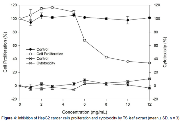
shows anti-proliferative activity and cytotoxic effects of the extract. The median effective dose (EC50) for inhibition of HepG2 cell proliferation was 7.45 ± 0.37 mg/mL.
Induction of apoptosis by TS leaf extract
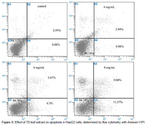
Pro-apoptotic effect is regarded as the most important route for anti-proliferative action of phytochemicals toward cancer cells [23]. The apoptosis effect of TS leaf extract was assayed by flow cytometric analysis and the results of annexin V-FITC/PI staining assay are displayed in . It was observed that the extract exhibited apparent effect on the percentage of early apoptotic cells. HepG2 cells treated with the leaf extract had an increase in the ratio of early stage apoptosis cells compared to the blank group. Remarkable enhancement was observed in cells treated with 6 mg/mL and 8 mg/mL of the extract. However, the percentage of late apoptosis in HepG2 cells was minor. The inhibiting effect of the extract on HepG2 cells proliferation was demonstrated mainly in early stage, implying a potential value of TS leaf as anticancer remedy.
Discussion
This study was focused on antioxidant and anti-proliferation activities of TS leaves extract towards HepG2 liver cancer cell line. Quercitrin and rutin were identified as the main flavonoids. Quercitrin (glycosylated form of quercetin), is the most common flavonoid found in nature and has been reported in vegetables and fruits such as apples, grapes, onions, tea [24]. Quercitrin is a bioflavonoid with antioxidant properties and is more easily absorbed than other forms of quercetin [25]. It is protective against UVB irradiation-induced oxidative damage to skin [24]. Rutin is a kind of flavanol glycoside, richly present in black tea, apple skin peels and buckwheat. It has been reported that rutin possesses antioxidant properties, scavenges hydroxyl and superoxide radicals and inhibits lipid peroxidation [26]. Thus the presence of quercitrin and rutin indicate that TS leaf has significant nutrition and health benefits. In plant extracts antioxidant activity cannot be ascertained by a single method due to the complex reactivities of phytochemicals.
Three methods i.e. PSC, ORAC and CAA were used to evaluate antioxidant potential of TS leaves. PSC and ORAC assays incorporate fluorescent probe to monitor reaction. These methods are rapid, precise, reliable, reproducible, and sensitive; and can produce acceptable results comparable to common chemical assays [12]. The assays also take into account the bioavailability, uptake and metabolism of the extract [18].
HepG2 cell culture model is relatively fast, represents the complexity of biological systems and is cost-effective. TS extract had lower EC50 value and higher antioxidant activity in the no-wash protocol than in wash-PBS protocol. This is due to the fact that PBS affects extracellular antioxidant activity, thereby reducing the intracellular antioxidant activity. These data demonstrate that the phytochemicals in the extract were able to pass through the cell membrane. Liao et al [27] observed that the inhibitory 50 % concentration values in the DPPH radical-scavenging activity of TS leaf extracts was 0.209 mg/mL. Although the mechanism of different antioxidant activities vary, our results are nonetheless comparable to that of Liao et al [27]. This indicates that TS leaf extract inhibits oxidative stress.
In an interesting study, TS leaf extract significantly inhibited ovarian cancer at the G2 phase in an in vivo xenograft model with no significant toxicity [28]. Furthermore, it has been reported that water leaf extracts of TS s had no acute lethal effect at test dose in male and female mice [29]. Our data also showed that the leaf extract of TS had antitumor effect but no cytotoxic effect.
Conclusion
The findings of this study reveal that TS leaf extract is rich in flavonoids, bioactive compounds responsible for its cellular antioxidant capacity and antitumor effects. These results provide scientific basis for the use of TS as a folk medicine for treating patients with liver cancer. The safety and efficacy of TS leaf makes it a potential natural medicine for managing hepatocellular carcinoma.
Declarations
Acknowledgement
References
Archives


News Updates